100 %

john paul

President

Lorem ipsum dolor sit amet, consectetur adipiscing elit. Duis lacinia porttitor ipsum, at gravida nunc ultricies quis. Quisque congue leo est, a venenatis ex congue a. Vivamus magna est, fermentum sed fermentum dapibus.Lorem ipsum dolor sit amet, consectetur adipiscing elit. Duis lacinia porttitor ipsum, at gravida nunc ultricies quis. Quisque congue leo est, a venenatis ex congue a. Vivamus magna est, fermentum sed fermentum dapibus.Lorem ipsum dolor sit amet, consectetur adipiscing elit. Duis lacinia porttitor ipsum, at gravida nunc ultricies quis. Quisque congue leo est, a venenatis ex congue a. Vivamus magna est, fermentum sed fermentum dapibus.Lorem ipsum dolor sit amet, consectetur adipiscing elit. Duis lacinia porttitor ipsum, at gravida nunc ultricies quis. Quisque congue leo est, a venenatis ex congue a. Vivamus magna est, fermentum sed fermentum dapibus.

Selenia Dimensions 2D3D

selenia-dimensions-2d-3d

Breast Imaging for Today and Beyond.The Selenia Dimensions 2D system is the ideal choice for centers that want a flexible digital breast imaging platform with state-of-the-art image acquisition and display features.Digital Mammography and Breast Tomosynthesis from Hologic.Selenia Dimensions starts with technology similar to that of our celebrated Selenia full-field digital mammography system. With its large amorphous selenium detector, it too can “direct capture” conventional digital mammograms with the same extraordinary clarity you’ve come to expect from Selenia. And incorporating a tungsten tube means we can do it with even less patient radiation dose than before. But Selenia Dimensions takes you to the next step in detection: fast, low-dose, high-quality tomosynthesis images of the breast.

1561543396908.png

download brochure

Request For Quote

  • Overview
  • Features
  • Benefits
  • Technical Specifications

Overview

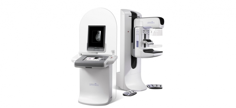
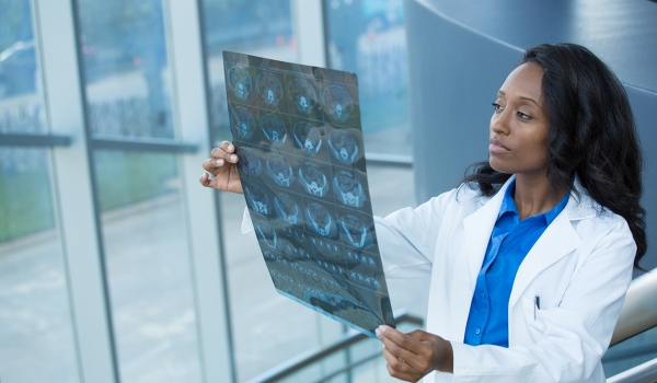
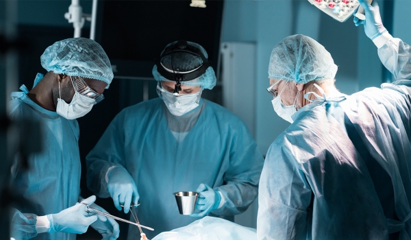
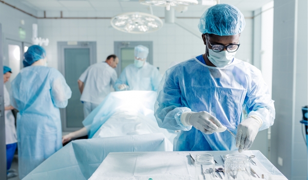
A flexible digital breast imaging platform providing state-of-the-art image acquisition and display

The Selenia® Dimensions™ 2D full-field digital mammography system, the newest addition to our family of breast imaging solutions, offers the superior image quality you have come to expect from Hologic and can be configured for 3D breast imaging when and if breast tomosynthesis is approved by the FDA.

The Selenia Dimensions 2D acquisition workstation features integrated x-ray control capabilities and an image acquisition console that offers a unique ergonomic design and an efficient user interface. An intuitive touchscreen display and an icon-driven user interface make it easy to use, improving patient throughput.

Images can be acquired and reviewed within seconds, and the imaging parameters can be adjusted quickly to meet the requirements of the examination. Several weeks of mammography examinations can stay locally on the acquisition workstation, to efficiently accommodate recalls.

Selenia Dimensions 2D has built-in quality controls which streamline system QC and assures optimal performance. Reject and repeat analysis reports are generated automatically, greatly simplifying administrative reporting. Altogether it’s a better way to capture, analyze and share information across the team.

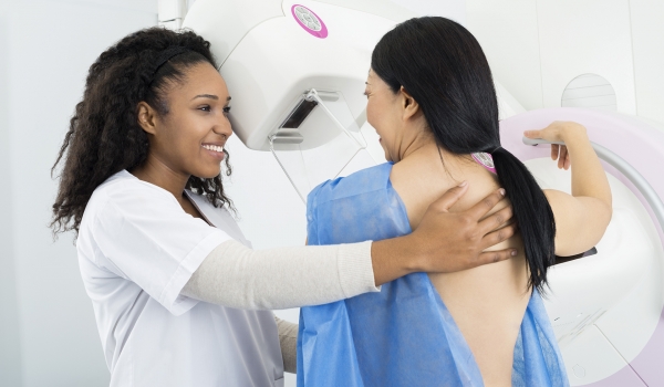
A Way to Be Surer. Sooner.

Breast tomosynthesis is the newly emerging method for delivering high quality images of breast tissue in three dimensions. We have good reason to believe it will be a quantum leap forward in breast cancer detection. Selenia Dimensions rapidly captures a series of low-dose images at different views around the breast.

From these original images, we can quickly compute a “synthetic tomogram” sequence, separating the full depth of tissue into discrete one-millimeter layers. This separation of tissue into virtual layers helps the physician clearly see features which might be obscured in a traditional mammogram.

Features

  • Proven Technologies

Direct Conversion Detector - Optimal Pixel Size

  • Preserves image sharpness by completely eliminating the image-degrading effects of light diffusion
  • 70 Micron pixel pitch, the most effective pixel size for visualization and characterization of smallest microcalcifications
  • Ongoing detector optimization program ensures enhanced stability and longevity

HTC Grid

  • Cross-hatch design reduces scatter in  X & Y direction
  • Allows 70-80% transmission of primary beam
  • Grid automatically retracts for magnification and tomo views
  • Goes from screening to diagnostic or tomo to 2D imaging in ~ 2 seconds
  • Grid never has to be handled, eliminating potential for damage

Optimized Tube Technology

  • Bi-angular tungsten (W) x-ray tube

                         Rhodium (Rh) filtration for most breast types 

                         Silver (Ag) filtration for breasts over 7 cm (compressed)

  • Tungsten dose: 1.0 - 1.2 mGy
  • W-Ag combination optimal for larger breasts*

                        Reduced dose without increasing exposure time

                        Decreases likelihood of patient motion

Innovative AEC

  • Intelligent tissue selection using two sensing areas for optimum technique selection
  • 15 cm travel from chest wall

                       Accommodates imaging of larger breasts

FAST Paddles

  • Provides proper and consistent compression across the entire breast without over-compression at the chest wall

                    Reduced motion artifacts

                    More uniform compression

                    Maximum patient comfort

Smart Paddle System

  • Smart Paddle System and fully automatic collimation allows small breasts to be imaged on large detector  
  • When paddle is shifted, AEC and collimation automatically moves to proper position

Optimized Detector always ready to acquire images

  • Latest, innovative selenium detector technology

                           Stable, long lasting detector

  • Ultrafast electronics for instant image read
  • Self-contained cooling system for independent temperature control
  • Independent power cycle for instant-on operation

High Patient Throughput without compromising patient care

  • High resolution preview image available 4 - 6 seconds following end of exposure
  • Rapid detector recycle time

                          Move from image to image without delay

  • Flexibility to accept, reject, or pend images

Ergonomic Design for enhanced patient comfort

  • Slim tubehead for easier, more comfortable positioning
  • 70 cm SID to facilitate interventional procedures
  • Easy access for wheelchair or stretcher
  • Recessed area on each side of C-arm for natural positioning of hands

Compression Paddle Innovations streamlined workflow

  • Lightweight, plastic-only paddles install directly on compression carriage; simply change plastic to move from paddle to paddle.
  • Each paddle registers through miniscule, surface-mounted electronics

Efficient Display

  • SecurViewDX 400 workstation optimized to view tomosynthesis images
  • Supports tomo, 2D, combined 2D+tomo, and tomo projections
  • Multiple display options to facilitate efficient review

Slices, stacks, cine, slabbing

  • Co-registered 2D + tomo display toggling
  • 2D CAD displayed on tomo slices

Networking Considerations

  • 100 Mbps may be acceptable, depending on overall traffic
  • 1 Gbps switched network is more than adequate for handling this volume of data
  • Web-based PACS clients may not be able to handle the transmission and display of tomo data

Benefits

Tomosynthesis Benefits

  • Removes overlapping structures
  • Increases lesion visibility
  • Facilitates margin analysis
  • Helps localize structures

Tomosynthesis Outcomes

  • Reduces recall rate (increased specificity)
  • Potential to increase cancer detection (increased sensitivity)

Image Resolution 

  • Reconstructed pixel size of 90µm is superior to original detector resolution  
  • Images are acquired at 140µm 
  • Resolution optimized for display on tomo-enabled workstations
  • Image quality proven through multi-center clinical trials

Technical Specifications

2D Imaging 

  • Delivers superb image quality at reduced dose 
  • Offers efficient workflow and a number of advanced features 

Tomo Imaging 

  • Can be used as an advanced diagnostic tool 
  • Offers superb diagnostic clarity at reduced dose 

Combo Imaging 

  • Designed to offer higher diagnostic accuracy in screening mammography 
  • Dose efficient option to maintain best image quality and workflow efficiency