100 %

Lorem ipsum dolor sit amet, consectetur adipiscing elit. Duis lacinia porttitor ipsum, at gravida nunc ultricies quis. Quisque congue leo est, a venenatis ex congue a. Vivamus magna est, fermentum sed fermentum dapibus.Lorem ipsum dolor sit amet, consectetur adipiscing elit. Duis lacinia porttitor ipsum, at gravida nunc ultricies quis. Quisque congue leo est, a venenatis ex congue a. Vivamus magna est, fermentum sed fermentum dapibus.Lorem ipsum dolor sit amet, consectetur adipiscing elit. Duis lacinia porttitor ipsum, at gravida nunc ultricies quis. Quisque congue leo est, a venenatis ex congue a. Vivamus magna est, fermentum sed fermentum dapibus.Lorem ipsum dolor sit amet, consectetur adipiscing elit. Duis lacinia porttitor ipsum, at gravida nunc ultricies quis. Quisque congue leo est, a venenatis ex congue a. Vivamus magna est, fermentum sed fermentum dapibus.

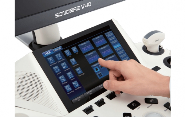
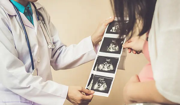
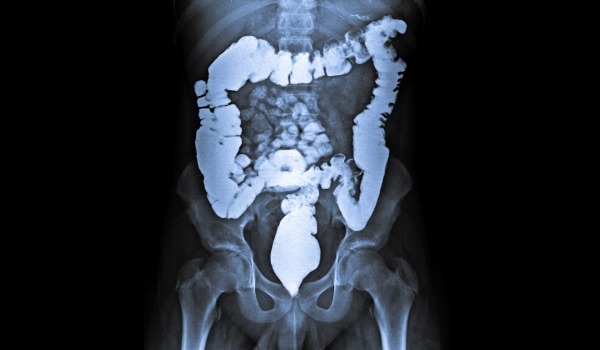
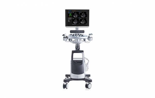
Ultrasound allows a medical examiner to examine the internal body structures such as tendons, muscles, joints, vessels and internal organs. The process is usually carried out with an intention to find the source of a disease or to exclude any pathology. This collected imaging insight can be used for either diagnosis or treatment (therapeutic procedures), as well as for guidance during procedures that require intervention, such as biopsies. Radiologists, cardiologists, or other medical specialists interpret the images and the required treatment will be carried out. Trivitron is associated with Hitachi Aloka for over 15 years – innovators of the ultrasound system, across the world.

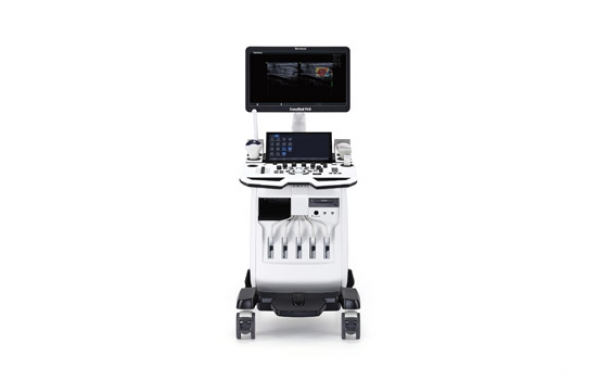
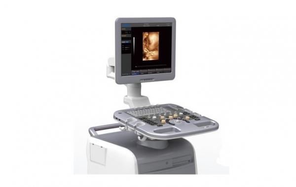
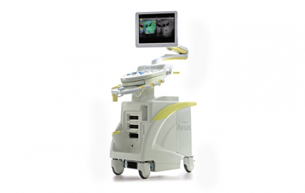
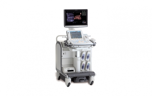
The Aloka Trivitron Ultrasound Imaging Series – SSD 9000, SSD 8000, and SSD 900 – redefines ultrasound imaging, delivering sharper images, quicker turnaround times, and greater confidence in clinical diagnosis. Each model embodies decades of imaging technology expertise and equips clinicians with the tools needed for accurate and efficient patient care.
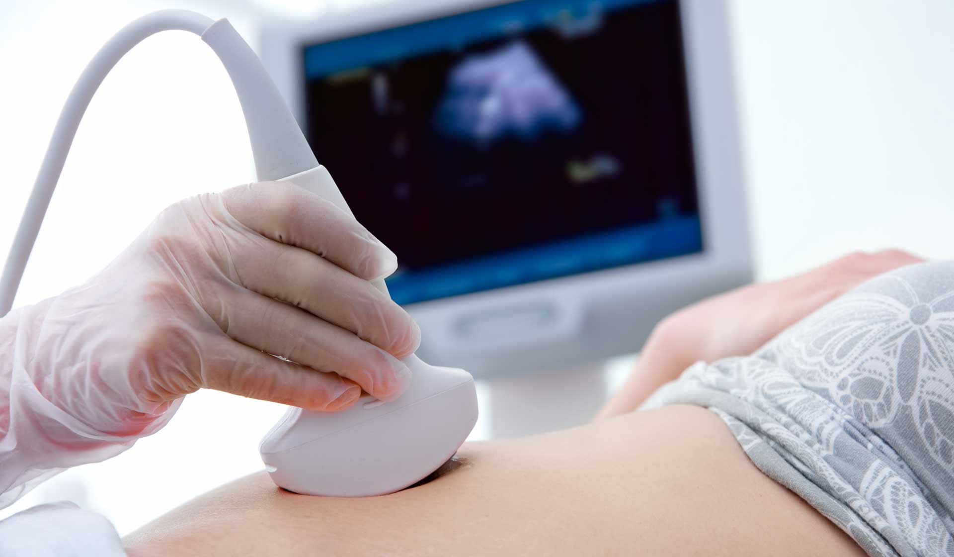
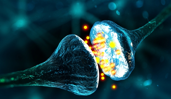
Doppler ultrasound depends on the Doppler Effect, a change in the wave's frequency that occurs from the motion of a reflector like a red blood cell. It is used to assess the flow of blood in a vessel, helping to determine the blood velocity and the obstructions in the blood pipes. It is often used to check the heart and the heartbeat of a growing fetus.
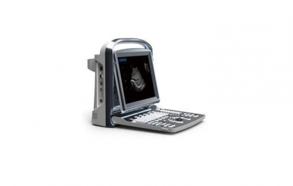
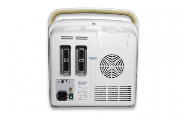
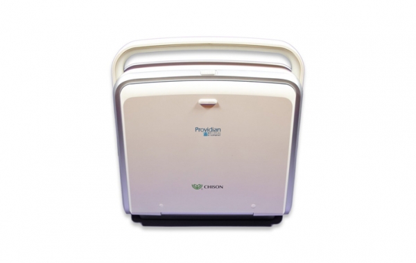
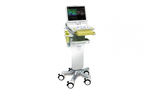
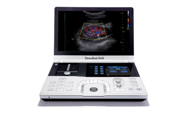
Chison Eco1 Exp is an inexpensive portable ultrasound with better imaging, full image, longer battery life. VET and Human calculations.

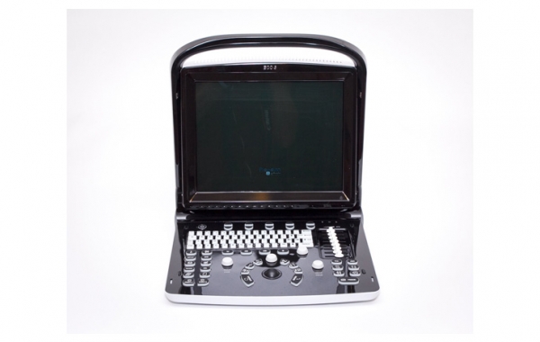
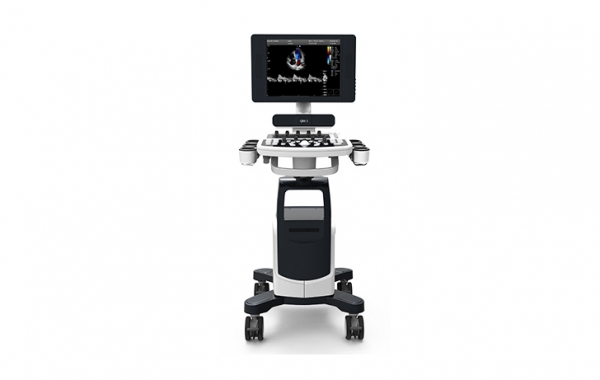
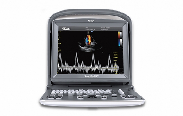
The new Chison Q5 ultrasound machine is a low-price portable color Doppler ultrasound machine. It is among the more versatile low-cost portable ultrasound machines with full measurement and analysis packages and presets for a wide variety of applications.


.jpg)